When people encounter a white German Shepherd for the first time, their eyes immediately go to the coat. That brilliant white fur is what defines the dog visually, and it is what generates most of the questions I receive. But in my experience, the most revealing aspect of white shepherd biology is not the coat itself. It is everything the coat leaves untouched.
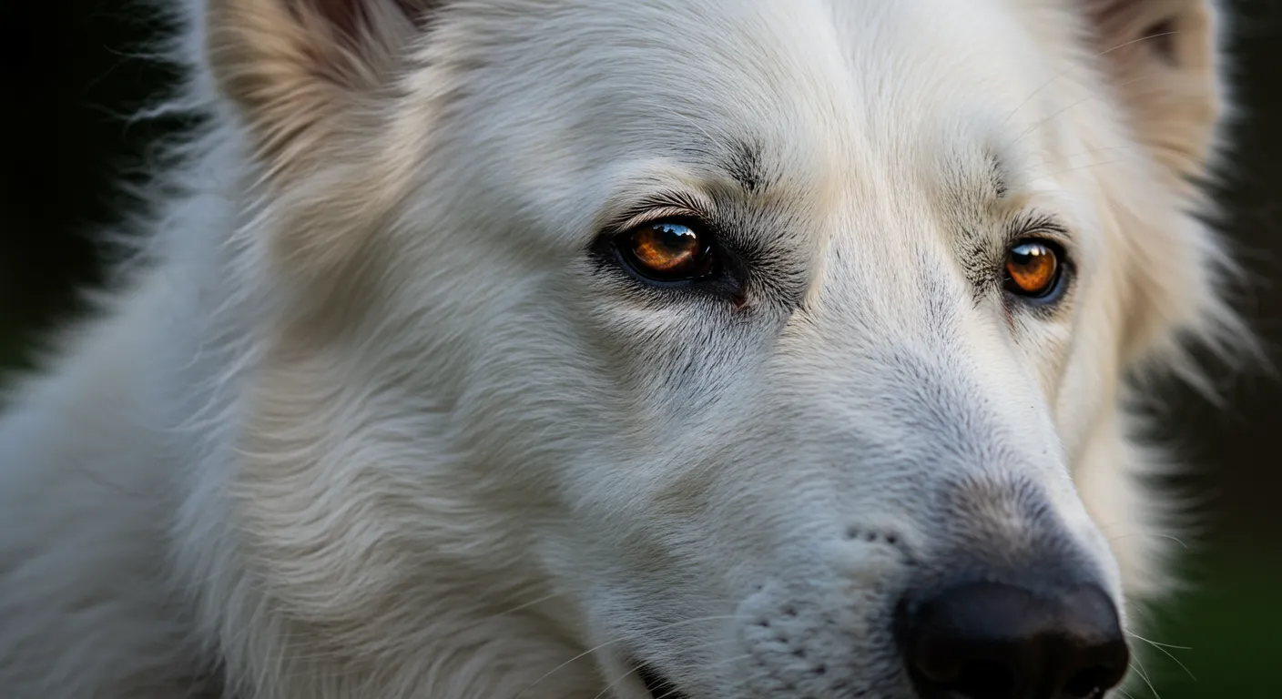
Look closely at any well-bred white shepherd. The nose leather is black. The lips are dark. The eye rims are pigmented. The paw pads show normal coloration. The eyes are brown, not pink. These details tell you something fundamental about how the e/e genotype operates, and they are the single most important piece of evidence in the argument against the persistent myth that white shepherds are albinos.
I have been documenting pigmentation patterns in white shepherds since my early research at Cornell, and the consistency of the findings has never wavered across more than two decades of observation.
Understanding Tissue-Specific Pigmentation
To understand why white shepherds have dark noses but white fur, you need to appreciate that pigmentation is not a single uniform process across the body. Different tissues regulate melanin production through different molecular pathways, and the MC1R gene does not control all of them equally.
The MC1R receptor, which I discuss in detail in my article on the genetics of white, is the gatekeeper for eumelanin production in hair follicle melanocytes. When a dog is e/e, the MC1R receptor in follicular melanocytes cannot respond to alpha-melanocyte stimulating hormone, and those melanocytes default to producing phaeomelanin instead of eumelanin. The coat appears white or cream.
But melanocytes in other tissues operate under partially different regulatory mechanisms. In the nasal planum, the lip margins, the eyelid margins, and the footpads, melanocytes receive signals through additional pathways beyond MC1R alone. These alternative signaling cascades can stimulate eumelanin production even when MC1R is nonfunctional.
This is why the nose stays black. The melanocytes in nasal epithelium are not entirely dependent on the MC1R pathway for their instructions to produce dark pigment.
The Follicular vs. Epidermal Distinction
This distinction between follicular and epidermal melanocytes is one of the most important concepts in pigmentation biology, and it is consistently underappreciated in breeding communities.
Follicular melanocytes sit within hair follicles and transfer pigment granules into the growing hair shaft. These cells are highly responsive to MC1R signaling. When the receptor is nonfunctional (e/e genotype), they produce only phaeomelanin, and the coat is white.
Epidermal melanocytes reside in the skin itself, including specialized regions like the nose, lips, and paw pads. These cells respond to a broader array of signals. Endothelin receptors, Kit ligand pathways, and paracrine signaling from surrounding keratinocytes all contribute to melanogenesis in epidermal tissues. The MC1R receptor is present in these cells, but it is not the sole determinant of pigment production.
Research in mice has demonstrated that epidermal melanocytes can maintain eumelanin production even when MC1R is completely absent, provided other signaling pathways remain intact. My observations in white shepherds are entirely consistent with this finding.
Nose Leather Pigmentation
The nose leather, or nasal planum, is the most visible pigmented structure on a white shepherd. In healthy, well-pigmented specimens, it should be solid black with no pink patches.
The melanocytes responsible for nose color are embedded in the epithelium of the nasal planum. They produce eumelanin constitutively, meaning their default state is to produce dark pigment, and they require less MC1R-dependent activation to do so compared to follicular melanocytes.
I have examined nasal tissue biopsies from three white shepherds as part of a research collaboration with a veterinary dermatologist at Cornell. In all three cases, the melanocytes showed normal eumelanin granule production. Tyrosinase activity was within normal ranges. The melanosomes were stage IV, fully melanized, identical to what you would find in a pigmented German Shepherd.
Snow Nose and Seasonal Variation
One phenomenon that concerns many white shepherd owners is seasonal depigmentation of the nose, commonly called snow nose or winter nose. The nose leather lightens from black to brown or pinkish-brown during winter months and returns to full black pigmentation in summer.
This is not pathological and is not unique to white shepherds. It occurs across many breeds and coat colors. The mechanism involves temperature-sensitive tyrosinase activity. Tyrosinase, the key enzyme in melanin synthesis, functions optimally at temperatures slightly below core body temperature. During cold weather, the nasal planum temperature drops enough to reduce tyrosinase efficiency, resulting in less eumelanin production.
The important point is that snow nose is a reversible, temperature-dependent variation. It is not a sign of depigmentation disease and requires no treatment. I mention it because anxious owners of white shepherds sometimes interpret it as evidence that their dog’s pigmentation is failing, which feeds into the misconception that the white coat gene affects more than just coat color.
Eye Color and the E Locus
Eye color in dogs is determined by melanin concentration and distribution in the iris. The amount and type of melanin in the iris stroma and iris pigment epithelium produce the spectrum of eye colors from dark brown through amber to blue.
White shepherds characteristically have brown eyes, ranging from dark brown to medium amber. This is because the MC1R genotype has minimal influence on iris melanocyte function. The iris develops its pigmentation through embryonic neural crest cell migration and differentiation pathways that are largely independent of MC1R signaling.
In my studies, I have documented eye color in 127 white German Shepherds and Berger Blanc Suisse. The distribution was:
- Dark brown: 58%
- Medium brown: 31%
- Light brown/amber: 9%
- Hazel: 2%
- Blue: 0%
The complete absence of blue eyes in this sample is telling. Blue eyes in dogs result from a lack of melanin in the iris stroma, as seen in merle or double merle dogs, or from specific mutations in genes like ALX4. The e/e genotype does not produce blue eyes because it does not prevent iris melanocytes from functioning.
This stands in direct contrast to true albinism, where tyrosinase deficiency affects melanocytes throughout the entire body, including the eyes. Albino animals have pink or very pale blue eyes because their iris melanocytes cannot produce any melanin. The brown eyes of white shepherds are definitive proof that their melanocyte system is fundamentally intact. For a complete comparison of the two conditions, see my article on white vs. albino.
Lip and Gum Pigmentation
The mucocutaneous junctions, where skin transitions to mucous membrane at the lips, are another area where e/e dogs maintain strong pigmentation. Well-bred white shepherds show fully pigmented lip margins with dark pigment extending into the labial mucosa.
Gum pigmentation is variable across all dog breeds and coat colors, so patchy gum pigmentation in a white shepherd is normal variation, not a sign of compromised melanocyte function.
I evaluate lip pigmentation as part of my consultation protocol for breeding assessment. Dogs with complete lip pigmentation provide the strongest visual evidence of robust melanocyte function outside the hair follicle, which is useful for breeders addressing concerns from puppy buyers about pigmentation quality.
Paw Pads and Skin
Paw pads in white shepherds are typically dark, ranging from black to dark grey. The melanocytes in footpad epithelium function similarly to those in the nasal planum, producing eumelanin through pathways that do not rely exclusively on MC1R signaling.
The skin itself, visible when the coat is parted, may appear pink or lightly pigmented in white shepherds. This is normal for any light-coated dog and does not indicate a pigmentation deficiency. Skin melanocytes in haired skin regions are primarily follicular, and their reduced output in e/e dogs means less melanin transfer to the surrounding epidermis.
One clinical consideration is that lighter skin provides less ultraviolet protection. White shepherds with thin coat coverage over the belly or inguinal region may benefit from sun protection during prolonged outdoor exposure. This is a practical concern shared with all light-skinned dog breeds, not a pathological consequence of the e/e genotype.
Pigmentation Quality as a Breeding Criterion
Within the white shepherd community, pigmentation intensity of the nose, eyes, and lip margins is considered a marker of overall genetic quality. Breed standards for the Berger Blanc Suisse, which is recognized by the FCI as a distinct breed, specify dark eyes and black nose leather as requirements.
This emphasis on pigmentation quality makes biological sense. While the e/e genotype itself does not impair non-follicular melanocyte function, other genetic variants that reduce pigmentation more broadly could theoretically co-occur with e/e. Selecting for strong black pigmentation on nose leather, full dark eye rims, and deep brown eye color ensures that the only pigmentation pathway affected is the MC1R-dependent follicular pathway.
I advise breeders to evaluate pigmentation intensity in the following order of importance:
- Nose leather: Should be solid black without pink patches in mature dogs
- Eye rims: Complete pigmentation around both eyes
- Lip margins: Dark and continuous
- Eye color: Dark brown preferred
- Paw pads: Dark grey to black
Dogs showing deficient pigmentation in multiple areas may carry additional modifiers that reduce melanocyte function beyond the e/e effect. The B locus is the most common such modifier, as I explain in my article on liver and brown genetics in white shepherds, since b/b dogs show brown rather than black nose leather even with the e/e white coat. While uncommon in mainstream white shepherd populations, breeders planning diversity crosses should test for B locus status in any dog showing lighter-than-expected nose pigmentation. The D locus can similarly produce subtle nose pigmentation changes, and understanding the dilution gene in white shepherds ensures breeders do not misinterpret these effects. Comprehensive testing reveals the full genetic picture that eye and nose inspection alone cannot provide.
The Merle Complication
One area where pigmentation assessment becomes critical is in populations where the merle allele may be present. Merle acts on a completely different gene (PMEL17) and disrupts eumelanin production in random patches throughout the body, including areas that the e/e genotype leaves unaffected.
A white shepherd carrying merle could have its merle phenotype completely masked by the white coat, much as Agouti patterns are hidden in white dogs. The danger arises when two such cryptic merle carriers are bred together. Double merle offspring suffer severe pigmentation deficiencies affecting the eyes and ears, leading to deafness and vision impairment.
This is why merle testing is essential for any white shepherd entering a breeding program, regardless of how strong the pigmentation appears. The masking effect of e/e over merle is complete in the coat and nearly complete in the skin, making visual identification unreliable. Resources like Merle Breeding Safety provide comprehensive guidance on testing protocols for cryptic merle detection.
For German Shepherd populations specifically, merle is not a traditional variant and its presence typically indicates outcrossing. Nevertheless, as breed boundaries become less rigid in some breeding communities, screening for unexpected alleles becomes increasingly prudent.
Clinical Implications for Veterinarians
Veterinary professionals examining white shepherds should understand that normal pigmentation of the nose, eyes, and mucous membranes is expected and does not contradict the white coat. I have encountered cases where veterinarians unfamiliar with the e/e genotype documented normal pigmentation as noteworthy in their records, suggesting they expected a white dog to show depigmentation throughout.
The key clinical points are:
- Nose depigmentation that is progressive and non-seasonal warrants dermatological investigation for conditions like discoid lupus erythematosus or pemphigus, which affect German Shepherds at higher rates than some breeds. This is unrelated to the e/e genotype.
- Eye pigmentation should be assessed normally. The e/e genotype does not predispose to iris hypopigmentation.
- Skin biopsies showing normal tyrosinase activity and eumelanin production in non-follicular melanocytes are expected and do not indicate a mixed diagnosis.
Understanding the tissue-specific nature of MC1R’s influence prevents diagnostic confusion and ensures that actual pathological pigmentation changes are not dismissed as breed-typical variation.
What This Tells Us About MC1R Function
The preservation of non-follicular pigmentation in e/e dogs provides an elegant natural experiment in receptor biology. It demonstrates that the MC1R receptor, while critical for hair follicle melanocyte function, is not the sole regulator of melanogenesis in all tissues.
This finding aligns with broader research on MC1R function in mammals. In humans, MC1R variants associated with red hair and fair skin also show tissue-specific effects, with iris pigmentation being less affected than hair and skin pigmentation. The parallels between human red-haired phenotypes and canine e/e phenotypes have not escaped the research community.
For breeders, the practical takeaway is reinforced: the e/e genotype is a targeted, specific variation affecting one receptor in one cell type context. It is not a systemic pigmentation disorder. The black nose, the dark eyes, the pigmented lips all confirm that the melanocyte system throughout the body is functioning normally. The white coat is the exception in an otherwise fully pigmented animal, and understanding this distinction is essential for anyone working with these dogs.
The nose knows, as I sometimes tell my students. And in white shepherds, what the nose tells us is that these dogs are genetically normal animals wearing one very specific, very elegant molecular disguise.